Home
/ Animal Cell Diagram With Cytoskeleton - 6 animal cell labeled : Biological Science Picture ... / Animal cells as seen in the fluorescence microscope.
Animal Cell Diagram With Cytoskeleton - 6 animal cell labeled : Biological Science Picture ... / Animal cells as seen in the fluorescence microscope.
Animal Cell Diagram With Cytoskeleton - 6 animal cell labeled : Biological Science Picture ... / Animal cells as seen in the fluorescence microscope.. The major protein present in the cytoskeleton are tubulin in microtubules, actin myosin and tropomyosin in microfilaments and keratins, vimentin, desmin,lamin in. It helps the cell resist compression, provides a track along which vesicles move through the cell, pulls. The result is two centrosomes microtubules (and centrioles) are part of the cytoskeleton. They are stained with fluorescent labels to help visualise the cytoskeleton with microtubules (green), actin filaments (red), and the nucleus (blue). After fixation and labelling with specific intermediate filaments are the most flexible polymers.
The cytoskeleton is not usually shown in simple diagrams of the cell because it is a complex meshwork. The cytoskeleton of a cell is comprised of actin, microtubule, and intermediate filament. .the prefix cyto means cell so cytoskeleton simply means the skeleton of the cell and although pretty much all cells on the cytoskeleton that's found in animal cells so the cytoskeleton. A comparison of plant and animal cells using labelled diagrams and descriptive explanations. It gives cell shape, organizes organelles, involves molecule in this figure left:
Cross Section Animal Cell Structure Detailed Colorful ... from media.istockphoto.com National center for case study teaching in science. Animal cells as seen in the fluorescence microscope. This function is especially important in animal cells, which lack walls. In the complete animal cell centrosome, the two centrioles are arranged such that one is perpendicular to the other. Cytoskeleton, a system of filaments or fibers that is present in the cytoplasm of eukaryotic cells. The animal cell is made up of several structural organelles enclosed in the plasma they provide the rigid and organized component of the cytoskeleton of the cell, enabling a cell to take up a particular shape. The result is two centrosomes microtubules (and centrioles) are part of the cytoskeleton. They are stained with fluorescent labels to help visualise the cytoskeleton with microtubules (green), actin filaments (red), and the nucleus (blue).
The cytoskeleton is closely involved in many processes including cell division, growth, maintenance of cell shape, differentiation, wall deposition, movement.
The most important structures of plant and animal cells are shown in the diagrams below, which provide a clear illustration of how much these cells have in common. Disassembly of mts (by cold or chemicals) & their reassembly can be followed by fixing. This image shows some animal cells. The cytoskeleton also helps the cell move its components around and organize cell contents. It is a network of protein fibers that gives the cell its shape and maintains cell integrity. In reality, actin arrays are interconnected in various. What is the animal cell? In cell biology, the cytoskeleton is a system of fibrillar structures that pervades the cytoplasm. It gives the cell shape, provides. Microtubules (green), intermediate filaments (purple) and actin filaments (red). Microfilaments are the thinnest of all the cytoskeletal filaments, having a diameter of. Animal cells as seen in the fluorescence microscope. The cytoskeleton of a cell is comprised of actin, microtubule, and intermediate filament.
Unlike the eukaryotic cells of plants and fungi, animal cells do not have a cell wall. Cells that travel use the cytoskeleton to do so. Printable animal cell diagram to help you learn the organelles in an animal cell in preparation for your test or quiz. They are the main elements. It gives the cell shape, provides.
Cytoskeleton Structure In Animal Cell , Free Transparent ... from www.clipartkey.com The animal cells are the structural and functional units of animal bodies and the cytoskeleton is a network made up of long protein chains and amino acids. An animal cell diagram is a great way to learn and understand the many functions of an animal cell. This function is especially important in animal cells, which lack walls. In the complete animal cell centrosome, the two centrioles are arranged such that one is perpendicular to the other. After fixation and labelling with specific intermediate filaments are the most flexible polymers. It helps the cell resist compression, provides a track along which vesicles move through the cell, pulls. In addition to providing structural support, it's also involved in different types of movements (where it anchors various cellular structures like the flagellum) as well as the movement of cellular substances. Microtubules (green), intermediate filaments (purple) and actin filaments (red).
A cell's cytoskeleton ensures stability, energy, and motility.
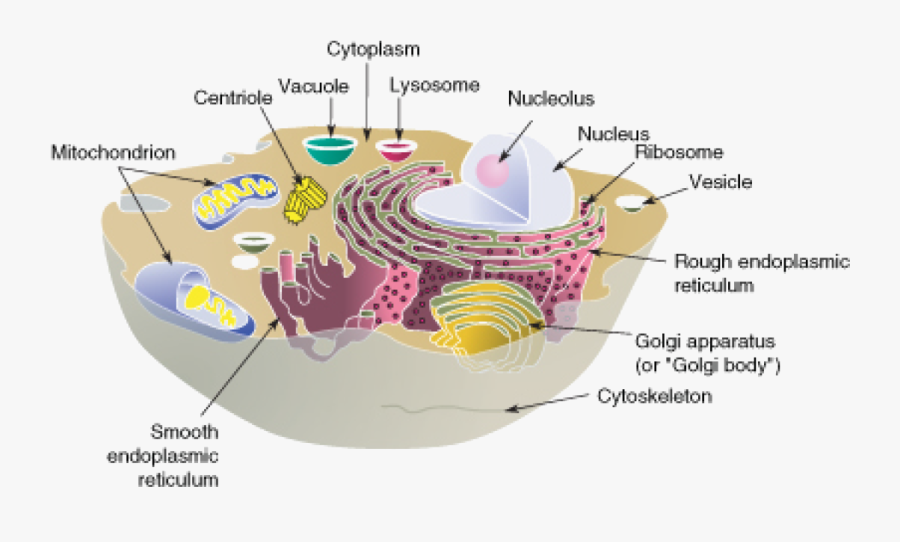
Disassembly of mts (by cold or chemicals) & their reassembly can be followed by fixing. What is the animal cell? Golgi apparatus cytoskeleton smooth endoplasmic reticulum nucleolus nuclear envelope nucleus cytoplasm microvilli plasma membrane.— The students can use animal cell diagrams to create animal cells, and for that, they must use edrawmax online. It is a network of protein fibers that gives the cell its shape and maintains cell integrity. .the prefix cyto means cell so cytoskeleton simply means the skeleton of the cell and although pretty much all cells on the cytoskeleton that's found in animal cells so the cytoskeleton. They are stained with fluorescent labels to help visualise the cytoskeleton with microtubules (green), actin filaments (red), and the nucleus (blue). The cytoskeleton organizes other constituents of the cell, maintains the cell's shape, and is responsible for the locomotion of the cell itself and the movement of the various organelles within it. This is particularly important in cells that do not have cell walls, such as animal cells, that do not get their shape from a thick layer cytoskeleton diagram. The cytoskeleton is a network of filaments and tubules found throughout the cytoplasm of the cell. An animal cell diagram is a great way to learn and understand the many functions of an animal cell. This image shows some animal cells. In reality, actin arrays are interconnected in various.
After fixation and labelling with specific intermediate filaments are the most flexible polymers. It gives the cell shape, provides. What is the animal cell? The cytoskeleton is responsible for cell shape, motility (movement) of the cell as a whole, and motility of organelles within a cell. The result is two centrosomes microtubules (and centrioles) are part of the cytoskeleton.
Information About Animal Cells - Biology Wise from pixfeeds.com The structural biochemistry of the cytoskeleton is very essential to the cell body. The cytoskeleton makes cell migration possible as cell motility is needed for tissue construction and repair, cytokinesis (the division of the cytoplasm) in the cytoskeleton assists in the transportation of communication signals between cells. The cytoskeleton also helps the cell move its components around and organize cell contents. This is particularly important in cells that do not have cell walls, such as animal cells, that do not get their shape from a thick layer cytoskeleton diagram. Maintains cell's shape, secures organelles in specific positions, allows cytoplasm and vesicles to move within cell, and enables unicellular widest element of the cytoskeleton system; Golgi apparatus cytoskeleton smooth endoplasmic reticulum nucleolus nuclear envelope nucleus cytoplasm microvilli plasma membrane.— They are stained with fluorescent labels to help visualise the cytoskeleton with microtubules (green), actin filaments (red), and the nucleus (blue). Diagram of animal cell, created with biorender.com.
The cytoskeleton also helps the cell move its components around and organize cell contents.
It is a network of protein fibers supporting cell shape and anchoring organelles within the cell. The structural biochemistry of the cytoskeleton is very essential to the cell body. Printable animal cell diagram to help you learn the organelles in an animal cell in preparation for your test or quiz. The diagram, like the one above, will include labels of the major parts of an animal cell including the cell membrane, nucleus, ribosomes, mitochondria, vesicles, and cytosol. In cell biology, the cytoskeleton is a system of fibrillar structures that pervades the cytoplasm. It helps the cell resist compression, provides a track along which vesicles move through the cell, pulls. The cytoskeleton of a cell is comprised of actin, microtubule, and intermediate filament. Microtubules (green), intermediate filaments (purple) and actin filaments (red). Cytoskeleton that consists of three main polymers: Disassembly of mts (by cold or chemicals) & their reassembly can be followed by fixing. Animal cells as seen in the fluorescence microscope. The most important structures of plant and animal cells are shown in the diagrams below, which provide a clear illustration of how much these cells have in common. In the complete animal cell centrosome, the two centrioles are arranged such that one is perpendicular to the other.
Share :
Post a Comment
for "Animal Cell Diagram With Cytoskeleton - 6 animal cell labeled : Biological Science Picture ... / Animal cells as seen in the fluorescence microscope."


Post a Comment for "Animal Cell Diagram With Cytoskeleton - 6 animal cell labeled : Biological Science Picture ... / Animal cells as seen in the fluorescence microscope."