Labelled Diagram Plant Cell Under Light Microscope / Q5 Make Sketches Of Animal And Lido - (refer to box 7.1 on p.. Cell is the basic building blocks of all organisms. Cell images from wiki images and other clip art sources. Raise the substage condenser to its top position and open the iris diaphragm all the way. Stains interact with a specific part of the sample, turning it a different colour from its surroundings. Under the microscope, it shows many different parts.
The high resolving power makes the electron microscope a very important research tool in microbiology. Be sure to only use the fine focus at the highest setting. Animal cells under a light microscope. Raise the substage condenser to its top position and open the iris diaphragm all the way. Labelled diagrams of typical animal and plant cells with editable layers.
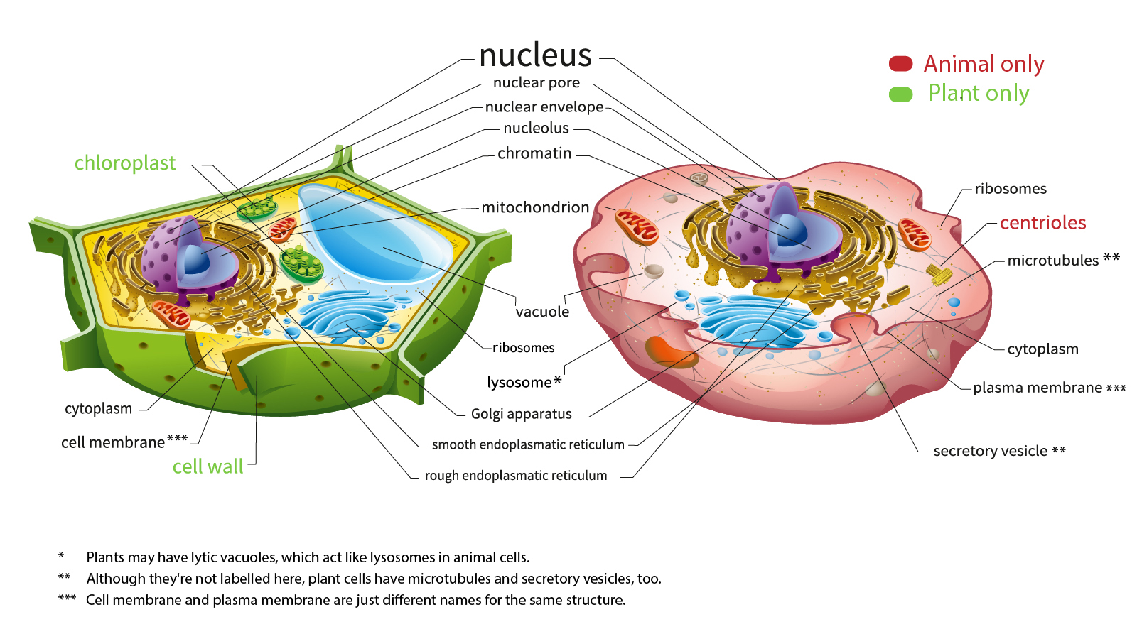
Structure Of Animal Cell And Plant Cell Under Microscope Diagrams Animal Cell Plant Cell Diagram Cell Diagram from i.pinimg.com Label the structures that you can see (e.g., cell wall repeat step 7 for higher settings of the microscope. Label the circle with the appropriate magnification. Learn about the size and function of plant and animal cells for gcse biology, aqa. The animal cell and plant cell diagrams are easily colorable, allowing students to differentiate the different parts of the cell quickly. Labeled diagram of plant cell, created with biorender.com. Elodea species or other freshwater aquarium plant. For example, iodine is often used to stain plant cells because it colours the starch. Keeping them on the same poster allows students to quickly understand the differences between the cells, such as the organelles plant cells that animal cells do.
Does anyone have a decent labelled diagram of a plant cell under an electron microscope?
For example, iodine is often used to stain plant cells because it colours the starch. A cell is a very tiny structure which exists in living bodies. Plug in the microscope and turn on the light source. 1 lab plant and animal cells, light microscopic analysis of leaf cross sections upper, structure of animal cell and plant cell under microscope, vacuole stock photos vacuole stock images alamy, cell structure teaching resources the science teacher. Transport proteins modified by the golgi body outside of the cell. Cell is a tiny structure and functional unit of a living organism containing draw a labelled diagram of animal cell and plant ncert. The high resolving power makes the electron microscope a very important research tool in microbiology. The typical characteristics that define the plant cell include cellulose, hemicellulose and pectin, plastids which play a major role in photosynthesis and storage of starch, large vacuoles responsible for regulating the cell turgor pressure. Plant cell science diagram clipart set includes: Observe the labeled diagram of plant. Light photomicrograph of helianthus stem cross section seen through microscope. A diagram of a plant cell. (refer to box 7.1 on p.
Plant cell science diagram clipart set includes: Once slides have been prepared, they can be examined under a microscope. Light photomicrograph of helianthus stem cross section seen through microscope. It is the small piece of cytoplasm contains a nucleus and covered by an outer protective. 1 lab plant and animal cells, light microscopic analysis of leaf cross sections upper, structure of animal cell and plant cell under microscope, vacuole stock photos vacuole stock images alamy, cell structure teaching resources the science teacher.
Here S How Plant And Animal Cells Are Different Howstuffworks from cdn.hswstatic.com The diagram is very clear, and labeled; Label the structures that you can see (e.g., cell wall repeat step 7 for higher settings of the microscope. Cell images from wiki images and other clip art sources. Be sure to only use the fine focus at the highest setting. When you look at animal or plant cells under the electron microscope, you can see a lot. Elements are high resolution 300 dpi png format with transparent backgrounds. Resolving power is the ability to distinguish between separate things which are close to each other. Transport proteins modified by the golgi body outside of the cell.
Cell is the basic building blocks of all organisms.
They can be observed easily in a phase contrast microscope under dark field. Plant cells contain many organelles such as ribosomes, the nucleus, the plasma membrane, the cell wall, mitochondria, and chloroplasts. Plant cell structure and parts explained with a labeled diagram. It is published by the american society of plant biologists. Stains interact with a specific part of the sample, turning it a different colour from its surroundings. For example, iodine is often used to stain plant cells because it colours the starch. Magnification, however, is not the most important issue in microscopy. When you look at animal or plant cells under the electron microscope, you can see a lot. Plant cell microscope image with labels cell theory plant cell. Learn the structure of animal cell and plant cell under light microscope. Light microscopes use a number of lenses to produce an image that can be viewed directly at the eyepiece. Juicy green plant cells under the microscope. Cell is the basic building blocks of all organisms.
Cell images from wiki images and other clip art sources. Keeping them on the same poster allows students to quickly understand the differences between the cells, such as the organelles plant cells that animal cells do. 1 2 applications and skills 1 2 2 practical 1 light microscopy. Light microscopes (also known as optical microscopes) are the original microscopes. A cell is a very tiny structure which exists in living bodies.
1 from Plug in the microscope and turn on the light source. The term 'cell' was coined to describe the small walled units that were observed in the sections they are just visible as small rods or spheres under light microscope. Juicy green plant cells under the microscope. Plant cell structure plant cell parts organelles and their. This shows a draw and label the structure of a generalized animal cell (i.e. Resolving power is the ability to distinguish between separate things which are close to each other. Limitations electron beams are deflected by air molecules, so the. Cell is the basic building blocks of all organisms.
Labeled diagram of plant cell, created with biorender.com.
Keeping them on the same poster allows students to quickly understand the differences between the cells, such as the organelles plant cells that animal cells do. Light photomicrograph of helianthus stem cross section seen through microscope. Plant cell science diagram clipart set includes: Once slides have been prepared, they can be examined under a microscope. Labeled diagram of plant cell, created with biorender.com. Plant cells are the basic unit and building blocks of life in organisms of the kingdom plantae. For example, iodine is often used to stain plant cells because it colours the starch. 129 of your text book for precise instructions on. Plug in the microscope and turn on the light source. Plant cells contain many organelles such as ribosomes, the nucleus, the plasma membrane, the cell wall, mitochondria, and chloroplasts. Learn the structure of animal cell and plant cell under light microscope. A cell is a very tiny structure which exists in living bodies. Let's go over the individual components of plant cells listed on a diagram such as the one above, and explore the roles that each of the organelles has.
Here's a photo of a plant cell under an electron microscope plant cell under light microscope. With light microscopy i can simply scrape some cells from my cheek smear them on a slide and look at them.
Share :
Post a Comment
for "Labelled Diagram Plant Cell Under Light Microscope / Q5 Make Sketches Of Animal And Lido - (refer to box 7.1 on p."


Post a Comment for "Labelled Diagram Plant Cell Under Light Microscope / Q5 Make Sketches Of Animal And Lido - (refer to box 7.1 on p."